
Project Coordinator: Assoc. Prof. Dr. Yeşim Serinağaoğlu Doğrusöz
Project Type: TUBITAK 1001 Scientific and Technological Research Projects Funding Program
Project Duration: 30 Months
Project Start Date:
Funded Personnel: 2 PhD Students (full-time), 1 MSc Student (full-time)
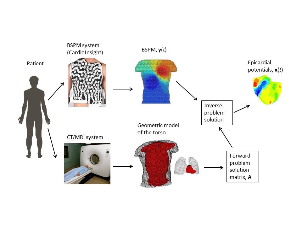
Conventional 12-lead electrocardiography (ECG), which is the primary method of diagnosing cardiac diseases, is widely used for detection of arrhythmias, but is insufficient to provide detailed distributions of electrical potentials, electrical conduction disturbances and sources of arrhythmia in the heart. More advanced diagnosis techniques are often invasive, their practice is at the very least uncomfortable to the patient, and can even lead to unwanted complications. One of the emerging diagnosis methods for heart arrhythmias is the noninvasive electrocardiographic imaging (ECGI). In this method, electrical potentials on the heart surface are estimated from body surface potentials (BSP) recorded with multi-channel ECG systems, and a mathematical model obtained using the geometrical structure of the body and the conductivity values of tissues. In particular, ECGI focuses on a type of arrhythmia referred to as premature ventricular contraction (PVC), in which electrical activity in the heart originates from a focal point in conflict with the normal conduction system. In advance stages, it can lead to fibrillation and even death. In clinical practice, treatment of these arrhythmias is performed by detecting PVC foci using invasive electrocardiographic imaging methods and ablation. Noninvasive ECGI has the potential for guiding the ablation procedure and shortening its duration by estimating the heart potential distributions and locating these foci before the procedure. However, in order for ECGI to become a clinically acceptable method, it is still necessary to develop new and innovative methods that are robust to uncertainties in the models, and to test these methods using clinical data.
Our aim is to obtain images of the cardiac electrical activity on the heart surface, and to detect PVC foci at clinically acceptable accuracy levels by using statistical estimation techniques. In these approaches, every parameter with uncertainty in their values (specifically epicardial potentials, measurement errors, geometric model errors, parameters used in their modeling, etc.) can be included in the model by means of a priori probability density functions (pdf), so that solutions can be obtained with higher accuracy than by using deterministic methods. However, the success of these methods highly depends on the use of correct models and a priori pdf’s. In this project, we will address some open questions in the literature, such as automatically determining a priori pdf’s from the available data, incorporating the uncertainties in the model parameters, and evaluating the accuracy of the results based on statistical metrics.